Kathleen Smith's Lab

banner

Images

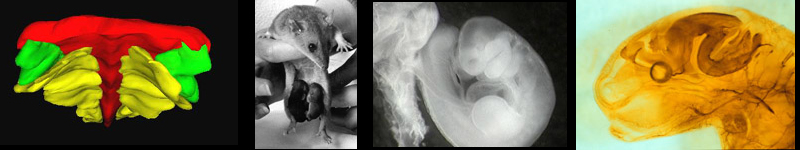
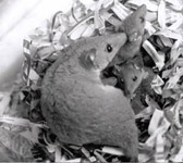
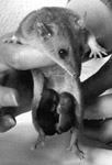
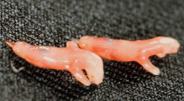
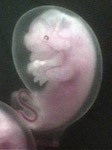
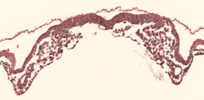
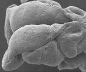
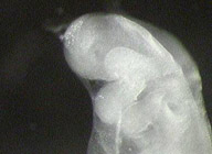
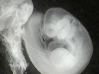
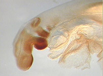
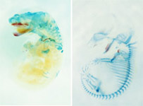
Below are images and captions to the figures shown on this webpage. In some cases the image is a link to a larger version. All images are © Kathleen Smith, and may not be used, except for educational purposes, without permission and proper attribution. All images of Monodelphis are Monodelphis domestica from the Smith breeding colony.

mother_with_young A female Monodelphis with young. These young are approximately 40-50 days old. They will be weaned at 50-60 days.
2_month_young A young Monodelphis, probably just after weaning.
Mother A female Monodelphis domestica with 4 young. These young are approximately 20-25 days old.
newborn_possum Newborn (less than an hour) Monodelphis young. These individuals are each about 1 cm long.
stage_33 A stage 32-33 Monodelphis embryo in its amniotic cavity. This individual is probably about one day before birth.
stage_29 A stage 29 Monodelphis embyo in situ in the uterus. At this stage the yolk sack placenta is just forming and the amniotic cavity is not fully formed.
stage_26_neural_tube A 3-dimensional reconstruction fo the neural tube of a stage 26 Monodelphis embryo. The reconstruction was from serial histological sections using Surfdriver software.
3d_reconstruction A 3-dimensional reconstruction of the neural plate (red), neural crest (green) and paraxial mesoderm (yellow) of a stage 23-24 Monodelphis embryo. The crest at this stage is sandwitched between the flat neural plate and the flat paraxial mesoderm. Reconstructed from serial sections using Surfdriver software. View is from the front
2h3_wholemount A head of a 2 day postnatal Monodelphis neonate stained with an antibody to neurofilament protein. Note the very large olfactory bulb and olfactory nerves.
stage_23_section A cross section of a stage 23-24 Monodelphis embryo (paraffin histology) in the region of the first arch. This section is from the series used in the 3-dimensional reconstruction above.
stage_25_sem SEM of a stage 25 Monodelphis embryo.
stage_26_embryo Photograph of a stage 26 Monodelphis embryo.
stage_29_embryo Photograph of a stage 29 Monodelphis embryo.
dll_wholemount A stage 26-27 embryo stained with an antibody to distalless antibody (G. Panganiban), showing migrating neural crest into the first and second arches.
skeletal_comparison Comparison of cleared and stained sections of a 2-day postnatal Monodelphis embryo and a day 14 embryonic Mus embryo, stained for cartilage and bone. This comparison reveals the steep anterior-posterior gradient in body axis development, and some of the most important differences in skeletal development.


back to top