
Figure 1. (Move mouse over the image to run the
animation)
Action-potential evoked calcium transient in a dendrite and
spines
[Home] [Publications] [Research] [People] [Positions] [Contact]
Imaging calcium-dependent signaling in neurons
Brain function relies on activity in networks of thousands of billions neurons wired through synapses. In the central nervous system, most synapses terminate on dendritic spines, tiny (volume 0.1-0.01 fL) mushroom-shaped protrusions emanating from the dendritic surface. These spines form the smallest known signaling compartments. Each spine contains a postsynaptic density (PSD), a specialized signal transduction complex, comprising hundreds of species of molecules, including synaptic receptors and channels, enzymes, and scaffolding proteins. Ca2+ dependent signaling via protein complexes associated with the PSD underlies synaptic plasticity and ultimately learning and memory. Our goal is to understand the signaling mechanisms in dendritic spines using novel biophysical techniques.
Regulation of voltage sensitive calcium channels in neurons
Voltage sensitive calcium channels constitute a major calcium source in dendrite, and play important role to connect neuronal activity to calcium dependent biochemical signaling in cells. Using two-photon-based Ca2+ imaging in combination with patch clamping techniques, we measured Ca2+ dynamics in individual spines in response to action potentials (Yasuda et al., 2004). This technique allows the measurement of the function and plasticity of voltage sensitive calcium channels with single molecule resolution. We found that a strong (but physiological) stimulus, such as a train of action potentials, can induce a pronounced depression of calcium channels over long times (>30 min). Surprisingly, depression of calcium channels abolished synaptic plasticity, revealing a coupling between Ca2+ channels and synaptic plasticity. The depression of Ca2+ channels was restricted to spines, and did not spread to their parent dendrites just ~ 1 μm away. In addition, in individual spines depression was all-or-none and occurred in a stochastic manner. This demonstrated for the first time that single spines function as independent biochemical reactors. Our analysis further revealed intricate signaling in spines. The depression pathway requires direct and local interactions between Ca2+ channels and protein kinases (CaMKII), providing strong evidence for the importance of Ca2+ microdomains in dendritic spines (Yasuda et al., 2003).
Figure 1. (Move mouse over the image to run the
animation)
Action-potential evoked calcium transient in a dendrite and
spines
Imaging protein-protein interactions
This work on calcium channels provides clues to one of the fundamental problems of Ca2+ signaling: how the single messenger can accomplish many diverse roles. The study suggested that the specificity comes from the local protein-protein interactions which activate distinct pools of enzymes. To gain a more detailed view of molecular interactions that play roles in specific signal transduction pathways, we have begun to develop an imaging technique based on FRET. FRET occurs when two fluorophores are within a few nanometers of each other. A part of the excitation energy from one fluorophore (donor) is transferred to the other fluorophore (acceptor), causing quenching of donor fluorescence and enhancement of acceptor fluorescence. Although the FRET effect is well known, it is notoriously difficult to measure FRET quantitatively in complex situations, such as inside neurons. We have used the measurement of fluorescence lifetimes, the time elapsed between fluorophore excitation and photon emission, as a measure of FRET: FRET decreases the mean lifetime of the excited state of the donor by removing the excited energy to the acceptor (reviewed in Yasuda, 2006). Unlike other methods, the measurement does not suffer from variable local concentration of donor and acceptor, or wavelength-dependent light scattering by tissues.
A number of obstacles need to be overcome to apply fluorescence lifetime measurements to post-synaptic signaling at the single synapse level. First, because often only a few copies of each protein exist in a spine, single-molecule sensitivity is required. Second, measurements need to be performed in highly light-scattering brain slices or even the intact brain. To overcome these difficulties, we have combined fluorescence lifetime measurements with two-photon microscopy and highly sensitive fluorescence detection (Two-photon Fluorescence Lifetime Imaging Microscopy or two-photon FLIM)(Svoboda and Yasuda, 2006; Yasuda R et al., 2006; Yasuda, 2006).
To measure fluorescence lifetimes we use a pulsed laser and measure the arrival times of photons at a photo-detector after a laser pulse. Because photon emission is a stochastic event, the histogram of the photon-arrival times is exponential. FRET between donor and acceptor causes the decay time constant of the donor to decrease. For mixed populations of fluorophores the lifetime distribution gives multiple exponential curves. It is relatively simple to deconvolve these curves to determine the relative population of donors that are bound to acceptors (Yasuda, 2006).
Imaging activity of Ras
One of the most critical signaling pathway for synaptic plasticity is Ras pathway (Zhu et al, 2002). Although the biochemical properties of Ras are well characterized, many mysteries remain about how Ras may be coupled to synaptic plasticity. Now, with two-photon FLIM, we can image dynamics of Ras activity to probe when and where Ras is activated to affect synaptic properties. To visualize Ras activity, we label Ras and Raf with GFP and RFP respectively. When Ras-GFP is activated, Raf-RFP binds to Ras-GFP leading to a robust FRET signal (Yasuda et al., 2006).
When 2-photon glutamate uncaging is used to stimulate a single spine, Ras activation occurs initially at the stimulated spine, and subsequently spreads more than 10um into the parent dendrite and nearby spines (Figure 2). The same stimulation induces both structural and functional plasticity in the stimulated spine, as indicated by a long-term (~60 min) spine enlargement associated with a potentiation of postsynaptic glutamate receptor current. To understand how Ras activation spreads and the role it may play in synaptic plasticity, we are now developing techniques to image molecules upstream and downstream of Ras.
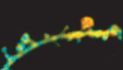
Figure 2. Imaging activity of Ras in individual spines. A single spine was stimulated by 2-photon glutamate uncaging. Redder the color is higher the Ras activity.
These methods are applicable to the measurement of activities of other enzymes and other signal-transduction pathways. By labeling the target proteins with a donor, and the peptide or antibody that binds specifically to the active protein with an acceptor, and then measuring FRET between these fluorophores, it will be possible to measure the activity of a broad class of kinases, phosphatases, channels and receptors. Furthermore, based on our work with calcium channels (Yasuda et al., 2003), we are also interested in probing protein-protein interactions between channels, receptors and enzymes in the synapse.
Studying biochemical signaling in cells has been difficult because one enzyme can play roles in many pathways, and likely only a subpopulation of the protein is activated in a specific pathway. Now, two-photon FLIM allows me to measure precise localization of enzymatic activities and protein-protein interactions in small subcellular compartments of living neurons in deep tissues. With this technique, we hopefully will be able to disentangle the complex signaling network in synapses.
References